How the Larynx (Voice Box) Works
Charles
R. Larson, PhD
If you love
opera, or if you admire the voices of pop singers such as Celine
Dion or Barbra Streisand, you may have wondered how it is these
marvelous singers are able to create such beautiful music with
this instrument we call the human voice. You may also know of
someone who has a bad voice or has had to have their voice box,
or larynx, removed because of illness or injury.
The
larynx is a critical organ of human speech and singing, and
it serves important biological functions as well. Let's have
a look at the larynx to understand its functions, what it
looks like and how it works.
It is
thought that the same factors that favored the evolution of
air-breathing animals on earth led to the evolution of the
larynx. Lungs are comprised of very delicate tissues that
must be maintained within strict biological limits, that is,
temperature, humidity and freedom from foreign particles.
Thus, along with the first air-breathing animals, there appeared
a primitive sort of larynx, whose one and only function was
protection of the lung. This function remains the most important
of those the larynx has assumed in subsequent evolutionary
developments. Now, of course we recognize that the larynx
is critical for human speech and singing. But we also should
realize that the larynx is important for swallowing, coughing,
vomiting and eliminating contents of the abdomen.
| If
you have ever felt your 'Adam's Apple', then you know
where the larynx is. The Adam's Apple is a protuberance
on the front of the larynx. This protuberance is part
of one of the main skeletal parts of the larynx, the thyroid
cartilage. The larynx is comprised of several other cartilages
as well as a single bone, the hyoid. Altogether, the cartilages
and bone provide a somewhat flexible and rigid framework
for support of softer tissues and muscles. |
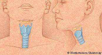 Figure
1: This figure illustrates the location of the larynx
within the neck. A: Hyoid bone, B: Thyroid cartilage,
C: Cricoid cartilage, D: Trachea. Click on the image for
a larger view.
Figure
1: This figure illustrates the location of the larynx
within the neck. A: Hyoid bone, B: Thyroid cartilage,
C: Cricoid cartilage, D: Trachea. Click on the image for
a larger view. |
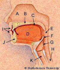
Figure
2: This figure depicts a section through the center of
the head and neck along the midline. In the top, one can
see the inside of the nasal cavity(A), oral cavity(C),
tongue(D), palate (B), jaw(J) and lips(I). Posterior to
the nasal and oral cavities lies the pharynx(E). In the
neck, the inside of the larynx(K) is shown anterior to
the pharynx and esophagus(H). Note the position of the
epiglottis(F) extending into the pharynx over the top
of the entry into the larynx from the pharynx (G). Click
on the image for a larger view. |
The
larynx is attached below to the trachea, or windpipe,
which goes down to the lungs in the chest and carries
the air we breathe. Immediately behind the larynx is the
pharynx. The pharynx is a tube-like structure that extends
from the upper border of the esophagus, which is at the
level of the bottom of the larynx, all the way up and
in back of the oral and nasal cavities. The upper border
of the larynx opens up into the pharynx. Thus, the air
we breath travels through the upper part of the pharynx
and then into the larynx. One unfortunate consequence
of human evolution is the fact that the food we eat and
the air we breathe share part of the pharynx, and if a
person tries to speak while swallowing food, the food
can enter the larynx and cause the person to choke. This
choking actually serves an important function, keeping
food out of or removing it from the larynx and trachea
for protection of the lungs. Another key protective feature
of the larynx is a rather floppy cartilage known as the
epiglottis. The epiglottis folds down over the entry into
the larynx during swallowing to help keep food out of
the larynx. |
| Now
let's examine what's inside the larynx and learn about
the structures that are involved in choking, holding one's
breath or the creation of the sounds of singing or speech,
which we call vocalization or phonation. The inside of
the bottom of the larynx is round and shaped like a cylinder.
As air ascends through the larynx, it encounters two folds
of tissue that extend out from the left and right sides
of the larynx. These are known as the vocal folds. The
vocal folds are also called the 'true vocal folds' because
immediately above the ventricle, there is a second set
of folds called the 'false vocal folds', or ventricular
folds.
These are called 'false' because the early doctors upon
looking down into a patient's throat with the aid of
a bent mirror, often mistook the 'false' folds for the
'true' folds. However, the function of the false vocal
folds is thought not to be nearly so critical for airway
protection or vocalization as are the true vocal folds.
|
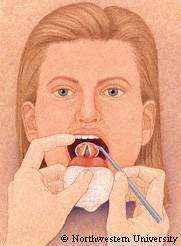
Figure
3: This figure illustrates how doctors visualize the larynx.
A small mirror attached to a bent rod is used to look
down into the larynx from the open mouth. Click on the
image for a larger view. |
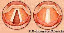
Figure
4: This figure illustrates two views of the vocal folds
observed from the laryngeal mirror (Figure 3). on the
left, the vocal folds are pulled laterally by the posterior
cricoarytenoide muscle to open the glottis, such as when
a person inhales, or takes a breath of air. On the right,
the vocal folds are pulled towards the midline by 'adductor'
muscles (see text), to close the glottis, as when as person
vocalizes or holds their breath. Click on the image for
a larger view. |
The
true vocal folds are attached to the inside of the thyroid
cartilage at about the level of the Adam's Apple. Posteriorly,
the vocal folds are attached to a set of cartilages known
as the arytenoid cartilages. There are several sets of
muscles that attach to the arytenoid cartilages, and by
their contraction, can move the arytenoids and along with
them the posterior part of the vocal folds. Because of
these anatomical relationships, the space in the middle
of the larynx between the vocal folds - the glottis -
is triangular in shape, with the narrow part of the 'V
' pointing towards the front. When the posterior cricoarytenoid
muscles contract, the arytenoid cartilages and vocal folds
are pulled laterally to open the glottis (abduction).
Every time we take a breath of air, we open the glottis
in this way. |
Most
of the other muscles pull on the arytenoids to either close
the glottis or stiffen the vocal folds. The lateral cricoarytenoid
and interarytenoid muscles pull the muscular processes of
the arytenoids and the vocal folds to the center of the glottis,
thus closing it (adduction). A similar muscle, the thyroarytenoid,
assists in closing the glottis and in addition makes the vocal
folds become very stiff. When all three of these muscles contract,
the glottis tightly closes. This is a configuration important
for swallowing, holding one's breath, or generating high abdominal
pressures associated with defecation, vomiting and child birth.
The last of the muscles to be considered here, the cricothyroid,
is on the front of the larynx and causes a rotational movement
between the thyroid and cricoid cartilages. Because the arytenoid
cartilages are attached to the back of the cricoid cartilage,
and the vocal folds are attached to the thyroid and arytenoid
cartilages, this rotational movement causes longitudinal stretching
of the vocal folds. This stretching is the primary means by
which we change the pitch of our voice.
The
muscular and cartilaginous actions mentioned above are similar
to those we use when we vocalize for speech or singing. In
order to vocalize, the edges of the vocal folds that face
the midline of the glottis are brought towards the midline,
but they are not pressed together tightly as they are when
we choke, swallow or hold our breath. Instead, the edges of
the folds are positioned so that they are lightly touching
each other or are close to each other. The process of vocalization
then results from blowing air up from the lungs past the edges
of the vocal folds. The flow of air initiates a repetitive
cycle of vibratory movements of the vocal folds. High air
pressure from below (subglottal pressure) blows the vocal
folds apart, and then elasticity of the vocal folds causes
them to return to the midline. Each time the folds are blown
apart, a small puff of air bursts up through the space between
the folds and 'excites' the air within the upper part of the
larynx and pharynx. Excitation is the term used to refer to
the generation of sound by the explosive quality of the burst
of air. This vibrational sequence of events repeats itself
on average about 110 times per second for an adult male and
about 200 times for an adult female. The sequence of these
air bursts takes on a tonal quality when repeated at high
frequency, and that is why our voices sound like a tone rather
than a series of mini explosions. When we sing, or raise the
pitch of our voices for other reasons, we contract the cricothyroid
muscles to stretch the vocal folds, and the stiffer the folds
become, the higher the frequency at which they vibrate.
Author
Charles
R. Larson, PhD
Professor, Departments of Communication Science Disorders and
Neurobiology and Physiology
Northwestern University |